Persistent Lameness
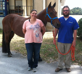
13-year-old Quarter Horse, Roanie, started showing signs of lameness in the spring of 2010. His owner, Jessica Niles, was concerned but always knew that his gait had never been completely normal. Roanie had an accident when he was a yearling that caused mild but permanent damage to right hindlimb.
“He has always shown hind-limb lameness because he has a large amount of scar tissue there,” Jessica said. “Pinpointing lameness in him has always been a challenge because it’s never just one thing, and it seems that one problem can lead to another and lead to another.”
Jessica called her primary care veterinarian, Dr. James Hilton, who performed an initial lameness evaluation. Based on the clinical signs, Dr. Hilton gave anti-inflammatories and recommended stall rest. When problems persisted, both Jessica and Dr. Hilton knew that the cause of the lameness was complex. Dr. Hilton referred Jessica to the UF Large Animal Hospital for a more thorough evaluation because of the many possible causes for the lameness.
One Visit, Many Options
When Jessica arrived at the UF Large Animal Hospital with Roanie, they were admitted to the Large Animal Surgery service and seen by Dr. Sarah Graham, Large Animal Surgery Specialist. With all horses exhibiting lameness, Dr. Graham began by performing a complete lameness examination.
“Roanie showed a right forelimb lameness grade 3/5 on the AAEP scale and a mild right hind limb lameness which we confirmed with the Lameness Locator,” Dr. Graham said. “The right hind limb lameness was secondary to his fibrotic myopathy because his gait was better at the trot than it was at the walk which is very characteristic of that condition. Since the fibrotic myopathy was mild, no treatment was recommended. Therefore, we were much more concerned about the right forelimb lameness and we proceeded with nerve blocks to try to isolate from where in the limb that the pain was coming.”
Roanie’s right forelimb lameness was resolved with a nerve block that numbed out the heel region. The next step was to radiograph the foot. Multiple radiographs of the foot revealed some changes in his navicular bone, but the changes were very mild and Roanie’s lameness was not. This meant that there was concern about other potential problems.
“The radiographs can show some bone changes but unfortunately the soft-tissues like tendons and ligaments will still be a mystery. An MRI has the ability to show much more, especially in a complex area like the foot,” said Dr. Graham. “Our high-field MRI is the most powerful diagnostic tool that we have available for horses and was Roanie’s best option for finding the cause of his lameness. Once we know what the specific cause of the problem is, we can begin an appropriate treatment and rehabilitation plan.”
In the same visit, an MRI of Roanie’s front feet was performed. The UF Large Animal Hospital’s Equine Lameness and Imaging team, including board-certified surgeons and radiologists, were able to determine that the lameness was caused by several abnormalities within the foot including navicular bursitis, navicular suspensory desmitis and tendonitis of the deep digital flexor tendon (DDFT) which are all soft-tissue injuries.
“The MRI showed a classic cluster of injuries within the foot that have often been referred to as Navicular Disease or Navicular Syndrome in the past,” Dr. Graham said. “The findings on the MRI were able to confirm our suspicions about Roanie’s lameness from the radiographs and lameness examination. Unfortunately, Quarter Horses seem to be predisposed to degeneration of the navicular bone and apparatus, which can lead to pain, lameness, decreased performance and early retirement. But, Roanie was early in the course of the disease and is very fortunate to have an owner like Jessica who was willing to give the tendon time to heal and consider some of the treatment options that we have available.”
Treatment & Rehabilitation Plans at UF
Treatment began right away. While Roanie was still under general anesthesia for the MRI, Dr. Graham and her colleagues performed a fluoroscopic-guided injection of the navicular bursa to reduce inflammation in the structure and reduce pain. Roanie was also treated with extracorporeal shockwave to the foot. This treatment can improve comfort and speed healing of ligaments and tendons. Jessica was given strict instructions to keep Roanie in a stall and was given a program of controlled hand walking.
Roanie was also given a new prescription for shoes and during the same visit, Roanie was re-shod by UF’s farrier, Jody Schaible, who works under the direction of the UF Large Animal Surgery service.

“Roanie had mediolateral imbalance and an improper angle of his hoof which was putting extra strain on his deep digital flexor tendon,” Jody said. “We shod both front feet in three degree wedges with a rolled toe. This type of shoe is commonly used for horses with navicular pain. The shoes were fit full through the heels and set back from the toe. This provides easier breakover and supports the heels. We also recommended that Jessica check his feet daily and use bell boots at all times to make sure the shoes weren’t accidentally pulled off.”
At Jessica’s request, long-acting acupuncture with vitamin B therapy was also instituted. Acupuncture may stimulate nerve fibers to release endorphins and other neurotransmitters which can modulate pain and have a calming effect.
“Roanie has always bucked a lot,” Jessica said. “The aqua acupuncture with B-vitamin really helped mellow him out in order for him be comfortable in the stall which was required as part of his rehabilitation plan.”
A Long Road for Roanie
Jessica quickly adjusted to the new routine of Roanie’s treatment plan. Hand walking and stall rest was all that Roanie was able to do during those first few months with the exception of farrier care and acupuncture therapy at the UF Large Animal Hospital every 4 to 6 weeks. These maintenance treatments at UF allowed Dr. Graham to monitor Roanie’s progress and ensure that every step was taken to help Roanie overcome his injury. He was also allowed to start a swimming program at a local rehabilitation center that helped him to get some exercise and maintain fitness and condition.
Roanie improved slowly over the next couple of months, and the right front lameness had almost resolved completely. Dr. Graham was concerned about his long-term prognosis because this can be a progressive and degenerative condition, and at 5 months post-MRI, Dr. Graham noticed something new that meant Roanie was not going to be returning to work soon.
“We starting seeing lameness in the left front – and that was a very bad sign,” Dr. Graham said. “We discussed the option for repeating the MRI to see what changes had occurred over the previous five months and Jessica opted to do that. She needed to know if Roanie had any chance at returning to work.”
The recheck MRI showed similar changes in the left front foot but also showed that the right front had improved.
“This was when we discussed possible navicular bursa surgery and stem cell therapy,” Dr. Graham said. “I also recommended that we change his shoeing to a Denoix Reverse shoe (made by Grand Circuit Products, LLC). This shoe has similar features to what he was in before, but the horses seem to respond to it better. Because it is a reverse shoe, breakover is even easier than in a regular shoe. It also allows more weight to be distributed onto the bars of the foot, therefore giving the heel region more support without putting pressure on the frog and navicular bone.”
Stem cells were obtained from Roanie’s bone marrow and sent to a specialized laboratory at the Marion duPont Scott Equine Medical Center, a campus of the Virginia-Maryland Regional College of Veterinary Medicine, for processing. After 30 days the cells were ready, and were planned to be injected into the navicular bursas at the time of the possible arthroscopic surgery. When Roanie returned for surgery, he looked much improved in the new shoes. Dr. Graham and Jessica discussed options, and Jessica wasn’t comfortable with the idea of surgery.
“I was concerned about Roanie’s tendency to build scar tissue, which might happen after surgery,” Jessica said. “Even a bug bite or small cut turns into a huge scar for him.”
Jessica elected to go forward with the stem cell injection but not the surgery, and when Roanie returned for his recheck four weeks later, Dr. Graham was pleasantly surprised at how good he looked and how much improvement he had made.
Since Roanie was so comfortable, Dr. Graham recommended an increase in the duration of Roanie’s walking under saddle sessions and to begin trotting him for short periods, gradually increasing the amount of work that he was doing. Two months after stem cell injections, Jessica was able to get Roanie up to a canter and start trail riding him. It was a long road but Jessica was determined to stick with the plan and prepare for improvement.
Back in the Saddle

A year after the initial diagnosis, Jessica reported that Roanie was maintaining his level of soundness despite a significant increase in exercise and workload. Roanie was being ridden almost daily and even went to two horse shows and competed well. The stem cell therapy and Denoix Reverse shoes seemed to have been the final boost in helping Roanie overcome his lameness.
“It is tough to explain why the stem cells seemed to help so much,” Dr. Graham said. “I don’t think that they necessarily ‘healed’ all of his injuries, but they may have provided some powerful anti-inflammatory and modulatory effects. There is so much that we still need to learn about regenerative therapies, how and when to use them. Roanie has a degenerative condition that will probably still worsen with time but he is doing remarkably well right now. Part of that success is probably due to the fact that we were able to treat the ‘whole’ horse by providing consistent foot care, medical treatment and rehabilitation therapy.”
Now, almost two years later, Roanie is back to his regular schedule and Jessica plans to be showing Roanie at full speed in the spring. His visits to UF are less frequent but the acupuncture and farrier care are still consistent. He has stem cells banked that are available if he ever has another injury or if the navicular bursas need to be retreated.
“It was tough but you really have to want it,” Jessica said. “I’m so happy that UF had all of the tools I needed to get Roanie back to work again. They have everything I need all in one place and it’s always a great experience to visit them.”
View all UF Large Animal Hospital Success Stories


