

Adrienne Hudson had her first encounter with colic in 1990 when her 23-year-old pony died from sand colic. It was then that Hudson decided to learn everything she could to prevent colic in any other horses she owned.
“Maximum turn-out, the least possible amount of grain, semi-annual dental care, and electrolytes when there are big temperature variations were some things I gave importance to,” Hudson said. “I generally feed flakes of hay or if I have to feed off of a round roll, I feed it in portions – never in a big round bale left out for them to gorge on.”
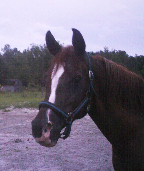
Hudson continued to learn more about colic prevention, preventive veterinary care and dietary maintenance and her horses stayed healthy. A few years later, Hudson purchased Rubiyat Kharma, or Ruby, when she was a weanling.
“Ruby was the culmination of my quest for a classic Arabian and I immediately had to take her home,” Hudson said.
Over the years, Ruby had some bouts of very mild colic when she traveled, but these were treated by her veterinarian with a dose of Banamine and she always recovered very quickly.
Immediate Veterinary Care
In April of 2012, however, Ruby was found cast in her stall. Hudson was able to get her up, but the mare continued to roll violently even when being walked. Hudson called her primary care veterinarian, Dr. Natalie Lamneck (UF College of Veterinary Medicine 2006), who arrived within 30 minutes and performed a rapid examination. Due to the severity of the colic signs, Dr. Lamneck told Hudson to take Ruby to the UF Large Animal Hospital for further care.
After Ruby arrived at UF, Dr. Chris Sanchez, Associate Professor of Large Animal Medicine, and Dr. Martha Mallicote, a large animal medicine resident, performed an emergency work-up. After finding blood in the abdominal fluid — an indicator of a compromised intestine — the veterinarians began to suspect a strangulating lesion of the small intestine. They recommended that Ruby go to surgery immediately.
Before taking over Ruby’s care, the large animal surgeon on duty, Dr. Sarah Graham, spoke with Hudson about the surgical procedures, expected outcomes and anticipated cost.
“I was a bit impatient to get Ruby to surgery,” Hudson said. “I’d made that decision before we left for UF, but Dr. Mallicote patiently and calmly reassured me that the operating room was being prepared as Dr. Graham and I spoke.”
Emergency Colic Surgeries at UF

Ruby was taken to UF’s specialized colic surgery operating suite where board certified veterinary surgeon, Dr. Sarah Graham, and large animal surgery resident, Dr. David Dymock, found a strangulating lipoma, or small fatty tumor, had cut off the blood supply to a large section of bowel. The veterinarians then surgically removed 25 feet of the jejunum, the middle section of the small intestine. The surgical procedure is called a “resection and anastomosis” and involves removing the damaged intestine followed by re-connecting the ends.
Through the use of a subjective system to decide when strangulated small intestine can be removed or left in the horse, Graham was able to determine how much of Ruby’s intestine needed to be removed. This system, pioneered by Dr. David Freeman, chief of large animal surgery, has led to higher long-term survival rates and lower complication rates.
Ruby’s surgical procedure was completed successfully and she recovered from anesthesia, but the following morning Ruby started showing signs of colic again. A nasogastric tube was passed into the stomach and excessive fluid was obtained — an indication that the small intestine was not working properly. The surgeons were concerned about the functionality of the anastomosis, as this is often the problem when horses develop reflux after that type of colic surgery. Since Ruby continued to reflux, Graham recommended that Ruby be taken back to surgery that day so that the anastomosis could be evaluated and repeated if necessary. This approach is consistent with the UF philosophy that emphasizes the importance of surgical treatment over aggressive or long-term medical management.
Ruby’s second surgical procedure revealed that a large blood clot in the mesentery, the tissue that connects the small intestine to the top of the abdomen, had caused a kink at the anastomosis site. The section of intestine affected by the blood clot, about 1.5 feet, was removed and the anastomosis was re-performed. While two colic surgeries within 24 to 48 hours is uncommon, the second surgical procedure was just as important as the first.
“In my mind, the repeat colic surgery was essential to the success of this case,” said Graham. “I don’t think that Ruby would have recovered or done as well in the long-term without it.”
In addition to fixing the anastomosis, the second surgery allowed the surgeons to repair a tear that had occurred in the body wall after recovery from the first surgery.
“We would not have taken the mare back to surgery to fix the body wall alone, but since we were already there we could address that problem as well,” Graham said. Large stent sutures were placed to support the closure of the body wall and prevent hernia formation.
According to Graham, some owners would have given up, but Adrienne didn’t.
“Her confidence in us and her belief in Ruby’s recovery really made a difference,” Graham said. “Taking Ruby back to surgery so quickly enabled us to save her owner money that might have been wasted on additional medications or a more prolonged hospital stay.”
“Dr. Graham’s efforts to help us contain the overall costs were greatly appreciated,” Hudson said. “She was never dismissive of our questions, concerns, suggestions or emotional attachment to our Ruby.”
A Positive Outcome for Ruby
Freeman emphasized the importance of the steps that contributed to Ruby’s successful outcome.
“Prompt referral is critical,” Freeman said. “Delays caused by continued therapy at home or at a hospital where the surgery cannot be performed might be well-intentioned, but can lead to a high failure rate.”
Luckily for Ruby, Hudson and her veterinarian acted quickly when the colic was noticed and the emergency colic surgery was performed at a nearby state-of-the-art facility by a board-certified veterinary surgeon and anesthesiologist. Other factors that contributed to Ruby’s success included: the owner’s awareness of colic; immediate referral for colic surgery by the UF large animal medicine team; the surgical techniques employed by UF’s large animal surgeons and the continuous support and care from the UF veterinary technical staff.
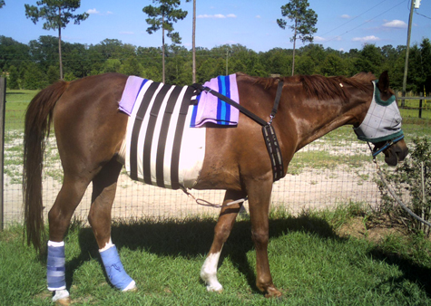
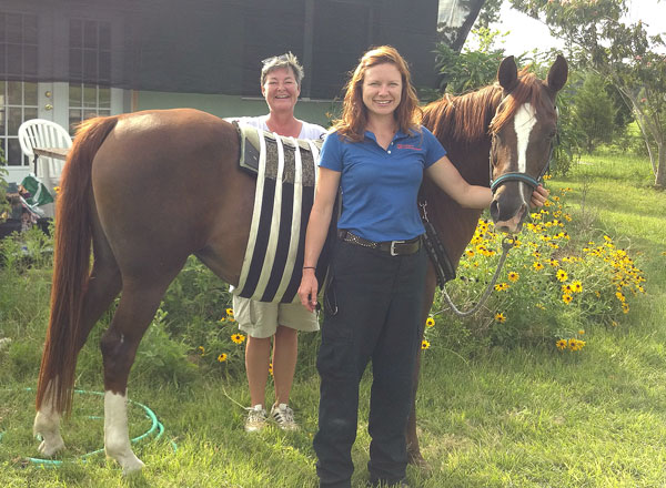
Just over a week after she first arrived for colic treatment, Ruby went home. She has shown no signs of colic since her second surgery. Veterinarians told Hudson that Ruby needed stall rest with hand grazing for two months before she could be released to a small paddock. Ruby’s diet increased gradually in the first week following surgery and after that she was allowed to eat her usual diet. Ruby used the CMTM Heal Hernia Belt (in photos) used as a post-surgical pressure bandage to support the incision and reduce the risk of forming a hernia.
“As far as the future goes for Ruby and us, I figure she’s good for another 15 or 20 years,” Hudson said. “Though I was trying my best to be clinical and unemotional at UF, the cocoon of understanding, caring and grace that surrounded Ruby and me was, for want of a better term, transcendent. I think that Ruby understands and appreciates everyone’s efforts to save her life.”
View all UF Large Animal Hospital Success Stories


